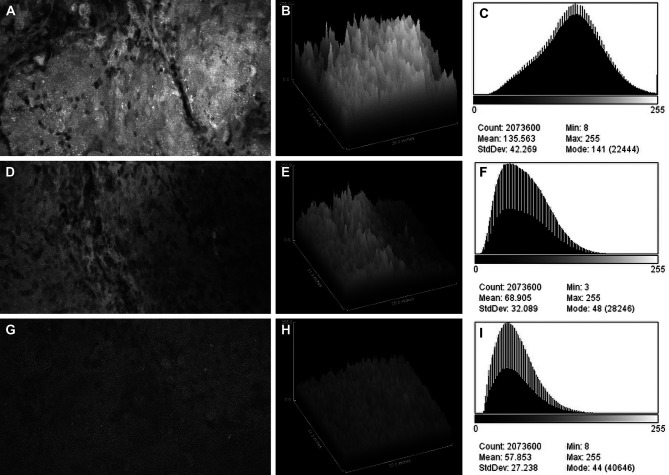
Figure 2.
Confocal laser endomicroscopy (CLE) images and data from (A–C) the fluorescein sodium redose, (D–F) initial-dose, and (G–I) single-dose groups. A CLE image (A, D, G) from each group is shown with a corresponding three-dimensional surface plot of pixel intensities (B, E, H) and a histogram of intensity values (C, F, I). Count, number of pixels; Mean, brightness; StdDev, contrast; Min and Max, minimum and maximum gray values within the image; Mode, most frequently occurring gray value within the selection corresponds to the highest peak in the histogram. Used with permission from Barrow Neurological Institute, Phoenix, Arizona.

