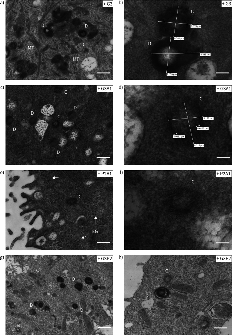
FIGURE 6.
High magnification images of the structures found in drug-treated reduced generation of multiple motile cilia (RGMC) cells. The different panels show representative high magnification images of accumulation of basal bodies “precursors” observed in RGMC cells after treatment with a, b) G3 (scale bar: 400 nm in a, 100 nm in b); c, d) G3A1 (scale bar: 400 nm in c, 50 nm in d); e, f) P2A1 (scale bar: 400 nm in e, 100 nm in f); and g, h) G3P2 (scale bar: 800 nm in g, 400 nm in h). Electron-dense deuterosomes were observed in high number and we observed several centrioles showing a “cartwheel” structure. Centriole size was ∼0.2 μm diameter (c–f [50]). None of these structures were observed in untreated controls. In (e), arrows indicate electron-dense granules. D: deuterosomes; C: centrioles; MT: microtubules agglomeration; EG: electron-dense granules.

