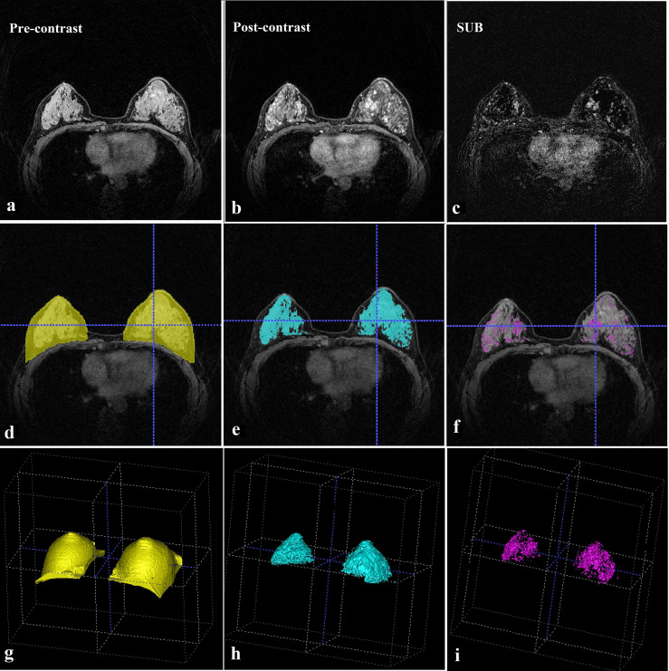
Figure 1.
MRI evaluation. (A) Pre-contrast breast MRI (T1-weighted fat-suppressed sequence). (B) This post-contrast (early) image corresponds to the T1-weighted image shown in (A). (C) Subtraction image from (A, B). (D) Whole breast segmentation (yellow). (E) Fibroglandular tissue segmentation (blue). (F) Enhanced fibroglandular tissue segmentation (purple). (G) Whole breast segmentation (3D). (H) Fibroglandular tissue segmentation (3D). (I) Enhanced fibroglandular tissue segmentation (3D). MRI = magnetic resonance imaging, 3D = three-dimensional.

