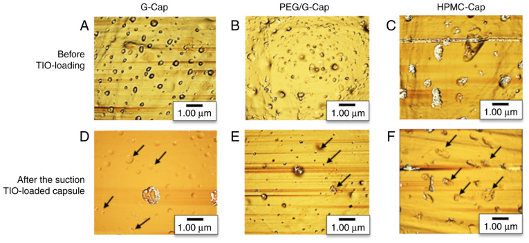
Figure 3.
Comparison of the capsule inner surface structure by atomic force microscopy. (A-C) Inner surface of each capsule before drug loading. (D-F) Inner surface of each capsule after drug release. Black arrows in (D-F) indicate the area where particles are placed in the valley. G-Cap, gelatin capsule; PEG/G-Cap, gelatin capsule containing 5% PEG4000 as plasticizer; HPMC-Cap, hydroxypropyl methylcellulose capsule; TIO, tiotropium bromide.

