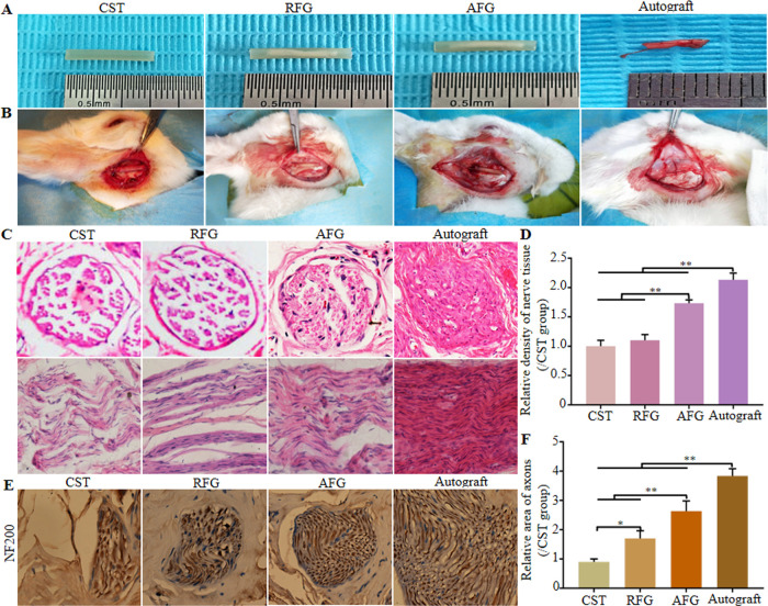
Figure 2.
Preparation and implantation of the different groups and histological observation. (A) Gross morphologies of implantation. (B) Graft implantation for bridging the 7 mm rabbit facial nerve defects: CST group: only empty chitosan conduit; RFG group: chitosan conduit filled with RFG; AFG group: chitosan conduit filled with AFG; Autograft group: autograft nerve that was rotated by 180°. (C) Hematoxylin–eosin staining of transverse and longitudinal sections of middle sections from the regenerative nerve at 12 weeks after surgery, and the relative density of transverse sections from the middle segment of regenerated nerve tissue (D). (E) Immunohistochemical staining (NF200, a marker for nerve fiber) of mid-segments from regenerated facial nerve at 12 weeks after surgery and the results of statistical analysis (F).

