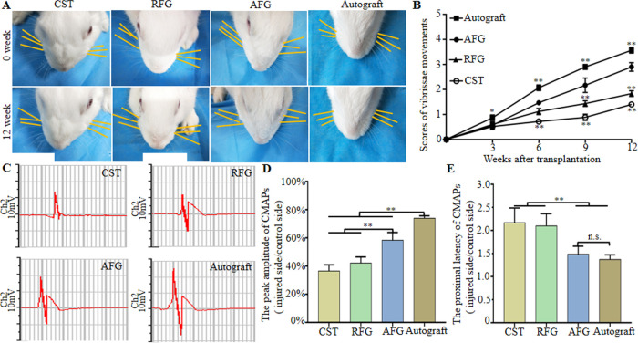
Figure 5.
Functional recovery of regenerated facial nerve. (A) Direction of the whiskers (as shown by the yellow line parallel to the corresponding beard for easy observation, and the right side of rabbit was operative side) in four groups at 1 and 12 weeks after surgery. (B) Score of vibrissae movement. (C) Electrophysiological evaluation of regenerated facial nerves 12 weeks after implantation and the statistical results (D) Peak amplitude of CMAPs (injury side/control side). (E) Proximal latency of CMAPs (injury side/control side). *P < 0.05, **P < 0.01 (n = 5).

