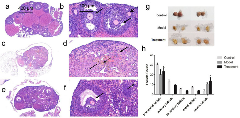
Fig. 4.
Ovarian tissue structure (a, c, and e: 50 × ; b, d, and f: 200 ×). a and b Preantral follicles (black dotted arrows) and antral follicles (thick black arrows) were visible in the young control group. c and d In the model control group, the nuclei were condensed (thick black arrow). In addition, a small number of infiltrating inflammatory cells (thin black arrow) and a small amount of brownish-yellow pigment deposition (black dotted arrow) were observed. e and f In the mUCMSC treatment group, more atretic follicles were observed, and the structures of the inferior antral follicles (thick black arrows) and atretic follicles (thin black arrows) were significantly improved. g Observation of ovaries from each group. h Numbers of follicles at various stages in each group; * P < 0.05 compared with the model group

