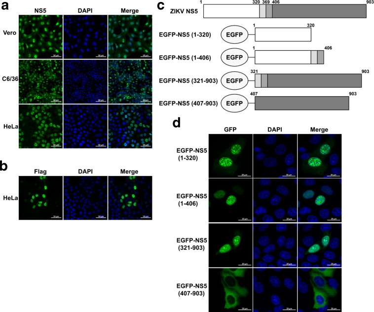
Fig. 1.
Both aa 1–320 and aa 321–903 of ZIKV NS5 accumulate in the cell nucleus. Vero, C6/36 and HeLa cells were infected with ZIKV at a m.o.i. of 1. At 24 h post-infection, cells were subjected to immunofluorescence staining for ZIKV NS5 (a). HeLa cells were seeded in 35 mm glass-bottom dishes and transfected with 1 µg NS5-WT plasmids. At 24 h post-transfection, anti-Flag antibody was used for immunostaining (b). Diagram of EGFP-tagged constructs (c). HeLa cells were seeded in 35 mm glass-bottom dishes and transfected with indicated EGFP-tagged NS5 constructs. At 24 h post-transfection, anti-GFP antibody was used for immunostaining (d). Images were captured by Zeiss LSM 880 CLSM by 63×oil immersion lens. The images used are representative of two independent experiments.

