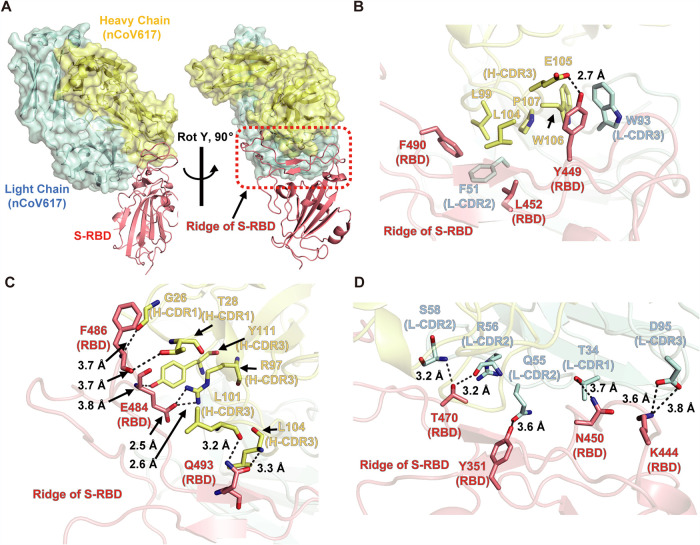
FIG 4.
Complex structure of MAb nCoV617 with SARS-CoV-2 S-RBD. (A) Overall structure of the MAb nCoV617-SARS-CoV-2 S-RBD complex with orthogonal angles. The light chain (cyan) and heavy chain (yellow) of MAb nCoV617 are illustrated with the ribbon and surface. SARS-CoV-2 S-RBD is illustrated with the ribbon, and the red dashed frame area is the ridge of RBD. (B) The Y449, L452, and F490 residues of S-RBD interact with the hydrophobic and strong hydrogen bonds of MAb nCoV617. (C) The hydrogen bond between the end of the S-RBD ridge region interacts with H-CDR1 and H-CDR3 of nCoV617 VH. (D) The long-distance hydrogen bonding interaction between S-RBD and the VL of nCoV617.

