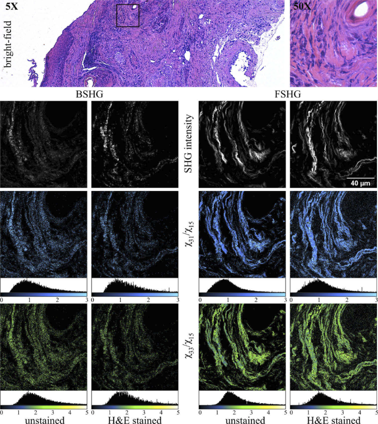
Fig. 2.
Complete image set obtained in the case of a skin tissue ROI. The square ROI in the BM image indicates the scanned area of 125 × 125 µm2. BSHG and FSHG images are acquired on both unstained and H&E-stained tissue sections. The SHG intensity image is the average of the 10 image PSHG set. χER images (χ31/χ15 and χ33/χ15) are computed for each case.

