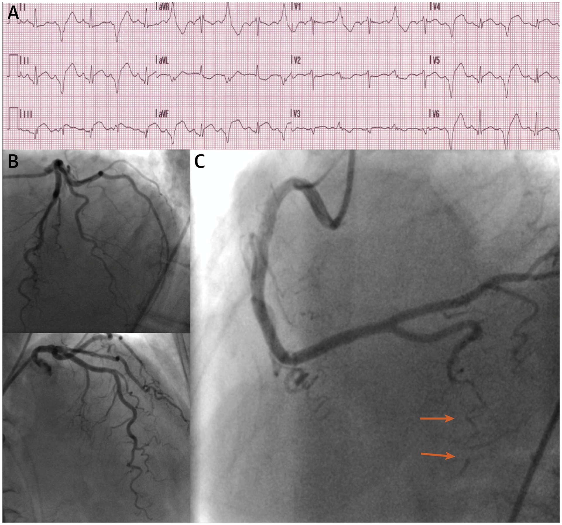
FIGURE 2. Myocardial Infarction With Nonobstructive Coronary Arteries of Case 1.

(A) Electrocardiogram showing an ST-segment elevation myocardial infarction. (B) Invasive coronary angiography (ICA) showing minimal coronary artery disease in the left anterior descending artery and left circumflex artery. (C) ICA showing a spontaneous coronary artery dissection of the right posterior descending artery, classified as a type 2B due to a long diffuse and smooth stenosis that extends to the distal tip of the artery (orange arrows).
