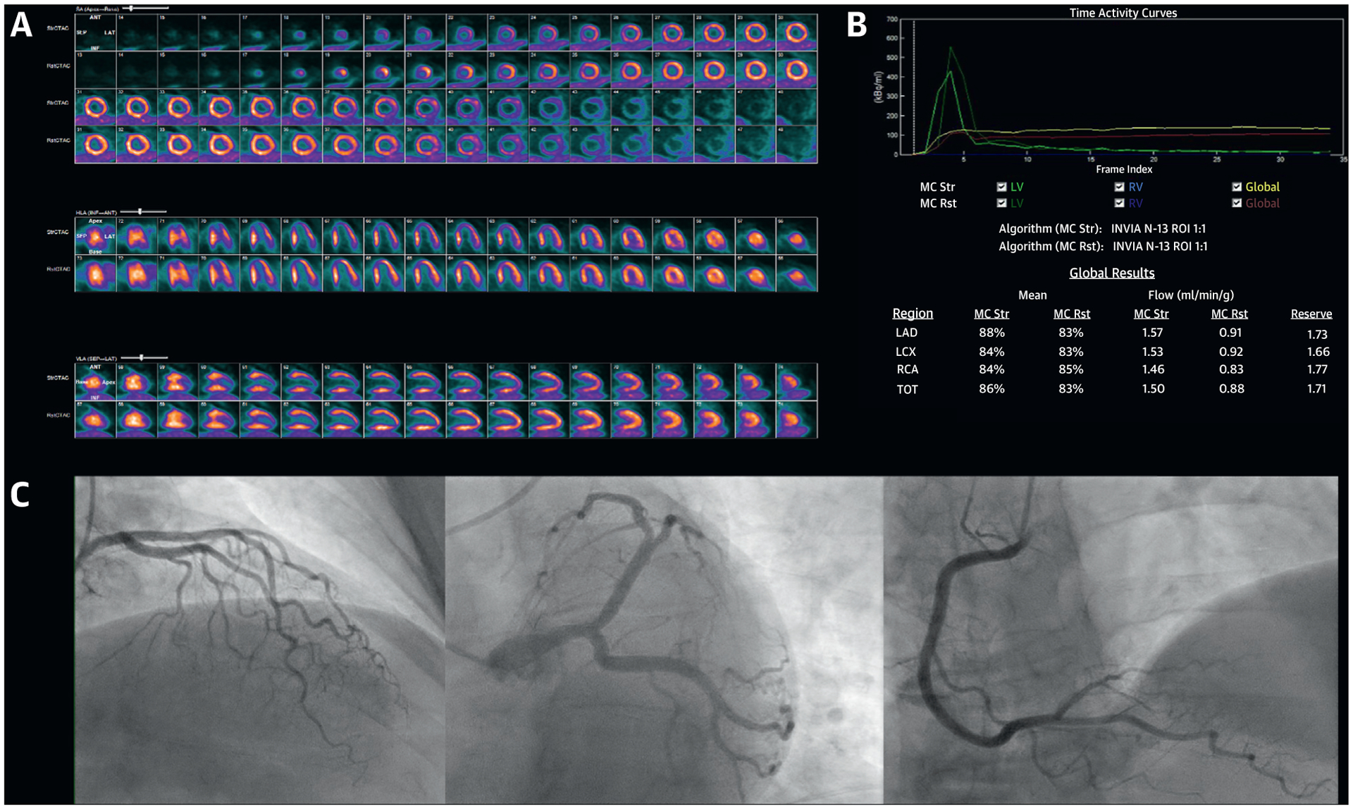
FIGURE 8. Integrated Imaging Findings of Case 5.

(A) Stress and rest 13N-NH3 myocardial perfusion positron emission tomography demonstrating severe ischemia involving the typical distribution of the mid to distal left anterior descending artery. (B) Positron emission tomography quantitative myocardial blood flow values demonstrating globally reduced coronary flow reserve. (C) Invasive coronary angiography demonstrating absence of obstructive epicardial coronary artery disease.
