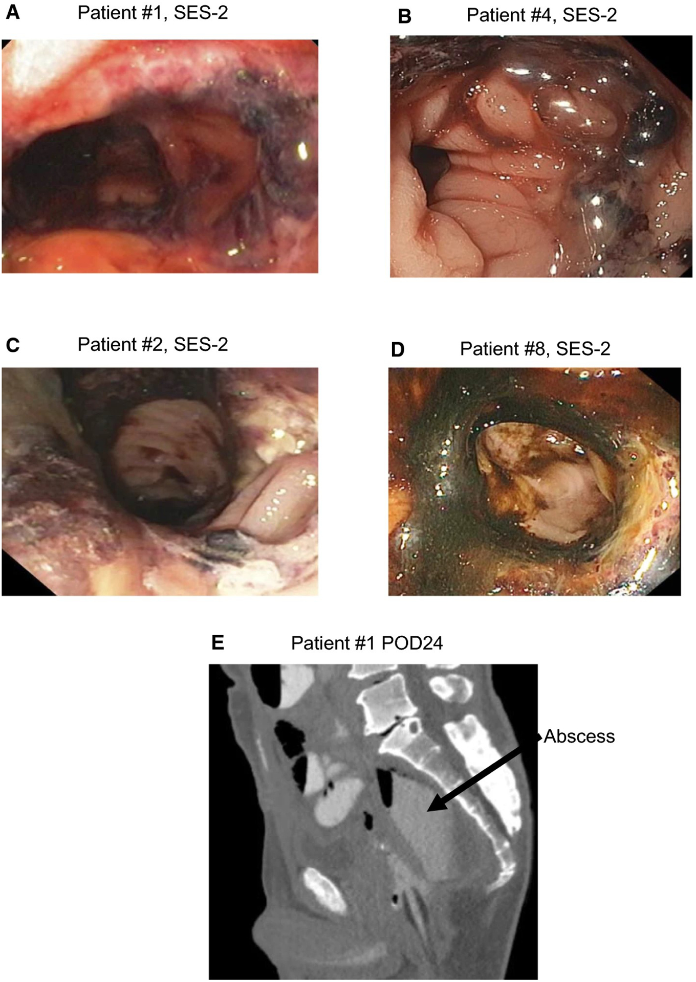
Fig.3. SES-2 endoscopy images demonstrating visible complications.

a, Edema on and extending the staple line in patient #1 at the SES-2 time point b, Edema on the staple line observed in patient #4 at SES-2; c, Ulceration observed on anastomotic line in patient #2 at SES-2; d, Edema on the staple line and ecchymosis patient #8 at SES-2; e, CT scan of patient #1 on postoperative day 24.
