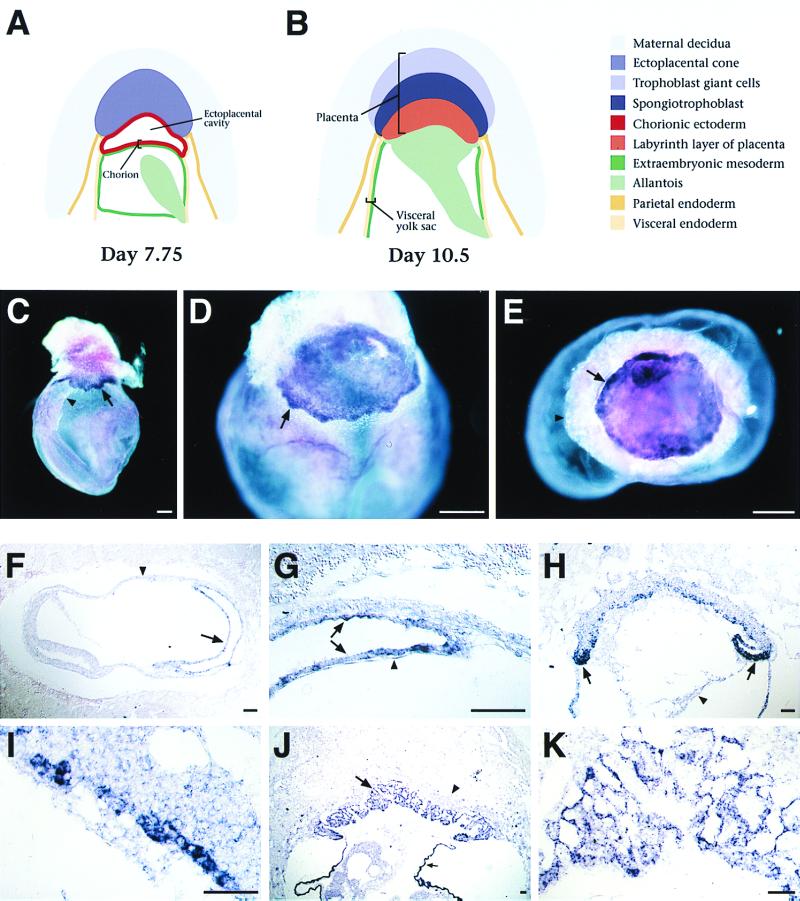
FIG. 2.
Esx1 expression in extraembryonic tissues. (A and B) Schematic depiction of stages in extraembryonic development using nomenclature from a previous report (24). (C to E) Whole-mount in situ hybridization analysis of intact egg cylinders. (C) Expression of Esx1 in the chorion (arrow) on day 8.0 (2 somites), just prior to fusion with the allantois (arrowhead). (D) Punctate staining in the developing placenta (arrow) on day 8.5, shortly after chorioallantoic fusion. (E) View of the day 8.5 placenta from below with the embryo removed; the ectoplacental cone and visceral yolk sac are beneath the plane of focus. Esx1 expression appears highest around the edges of the placenta (arrow). (F to K) Section in situ hybridization to cryosections of egg cylinders on days 7.5 through 9.5 of gestation. (F) Sagittal section through a day 7.5 egg cylinder (head-fold stage); the ectoplacental cone is to the right. Esx1 is expressed in the chorion (arrow) but not in the visceral yolk sac (arrowhead). (G) Higher-power view of panel F, showing staining of the chorionic ectoderm (arrows) but not of the chorionic mesoderm (arrowhead). (H) Sagittal section on day 8.5 of gestation, with scattered Esx1 expression in the newly formed placenta, with the highest levels at the inverted lateral margins (laminae) of the ectoplacental plate (arrows); no expression is observed in the allantois (arrowhead). (I) Higher-power view of the ectoplacental plate from panel H. (J) Low-power view of the placenta on day 9.5 of gestation. Expression is found in the developing labyrinth layer (arrow) but not in the spongiotrophoblast layer (arrowhead). Expression is also evident in the visceral endoderm of the yolk sac (small arrow); note that this is consistent with the expression of Esx1 during differentiation of ES embryoid bodies, which produce visceral endoderm. (K) Higher-power view of the labyrinth layer. In panels C to E, bars represent 200 μm; in panels F to K, bars represent 50 μm.

