
FIGURE 2.
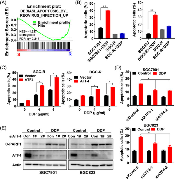
ATF4 stimulates drug‐induced apoptosis. (A) To assess the correlation of the expression of apoptosis‐related gene signature in resistant cells compared to sensitive cells, GSEA was used to analyse the gene expression profiles in chemoresistant cells (SGC‐R and BGC‐R) and their parental sensitive cells (SGC7901 and BGC823). (B) Apoptosis of SGC7901 and SGC‐R (left) or BGC823 and BGC‐R (right) under DDP (1.2 μg/ml) incubation for 24 h was measured by a PI/Annexin V double staining assay. (C) Apoptosis of SGC‐R (left) or BGC‐R (right) cells with ATF4 over‐expression and DDP treatment for 24 h was measured. (D) Apoptosis of SGC7901 (up) or BGC823 (down) cells with ATF4 knockdown and DDP treatment for 24 h was measured by a PI/Annexin V double staining assay. (E) The expression of apoptosis marker cleave‐PARP1 (C‐PARP1) and ATF4 in SGC7901 (left) or BGC823 (right) cells with ATF4 knockdown and DDP (1.2 μg/ml) treatment for 24 h was detected by western blotting