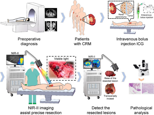
FIGURE 1.

Flow diagram of the study protocol. Patients first received preoperative imaging examinations to determine whether eligible for the study. On the day of surgery, after laparotomy, ICG with a dose of 0.5 mg/kg body weight was administrated intravenously before blocking the renal artery. Then, the tumour boundaries were marked on the kidney surface under the guidance of the NIR‐II images, which were followed by arteries clamping and tumour resection. After resection, images of the surgical margins and resected lesions in the white‐light illumination and NIR‐II region were acquired separately. Lastly, pathological examinations were carried out
