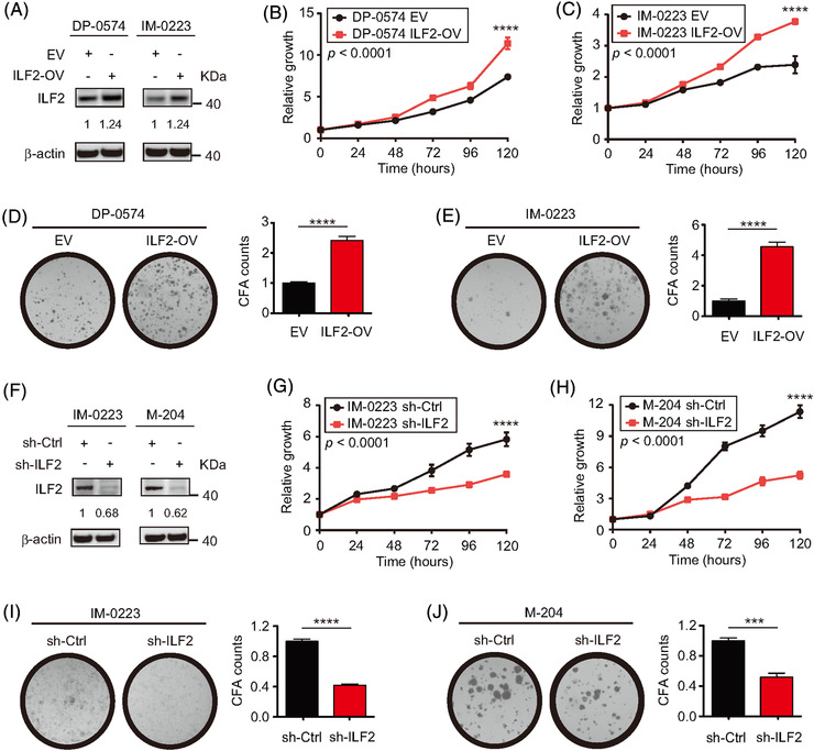
FIGURE 2.

ILF2 expression determines cell proliferation and colony formation ability in metastatic melanoma. (A) Western blot and quantification for ILF2 in two melanoma cell lines with ILF2 overexpression (ILF2‐OV). β‐actin was used as the loading sample control. (B and C) Proliferation assays in two melanoma cell lines with ILF2‐OV. (D and E) Representative colony formation images and quantification plots for the two melanoma cell lines with ILF2‐OV. (F) Western blot and quantification for ILF2 in two melanoma cell lines with ILF2 knockdown. β‐actin was used as the loading sample control. (G and H) Proliferation assays in two melanoma cell lines with ILF2 knockdown. (I and J) Representative colony formation images and quantification plots for the two melanoma cell lines with ILF2 knockdown. Data represent the mean ± SD. ***p < .001 and ****p < .0001
