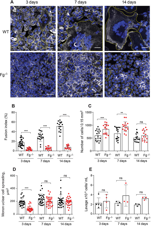
Figure 1. Fg−/− mice display reduced FBGC formation on biomaterials implanted intraperitoneally.
PCTFE sections were implanted in the peritoneum of WT and Fg−/− mice for 3, 7, and 14 days and analyzed by immunocytochemistry. (A) Explants were separated from the surrounding fibrous capsule, fixed, and incubated with Alexa Fluor 546-conjugated phalloidin (white) and DAPI (teal). Representative confocal images are shown. FBGCs are outlined (yellow). The scale bar is 20 μm. (B) Macrophage fusion was assessed as a fusion index, which determines the fraction of nuclei within FBGCs expressed as the percentage of the total nuclei counted. Five to six random 20× fields were used per sample to count nuclei. (C) The number of cells on the surface of explants retrieved at various time points was determined by counting nuclei in mononuclear cells and FBGCs. (D) Spreading of mononuclear macrophages on the surface of explants. Six to eight random 20x fields per sample were used to determine the cell area (5-6 cells/field). (E) The number of cells in lavage recovered from the mouse peritoneum before explantation. Results shown are mean ± S.D. of five independent experiments. n=6 (WT) and n=7 (Fg−/−) mice per each time point. For counting lavage cells 3-5 WT and 3-4 Fg−/− mice were used per each time point. Two-tailed t-test and Mann-Whitney U test were used to calculate significance. ns, not significant, **p < .01, ***p < .001.

