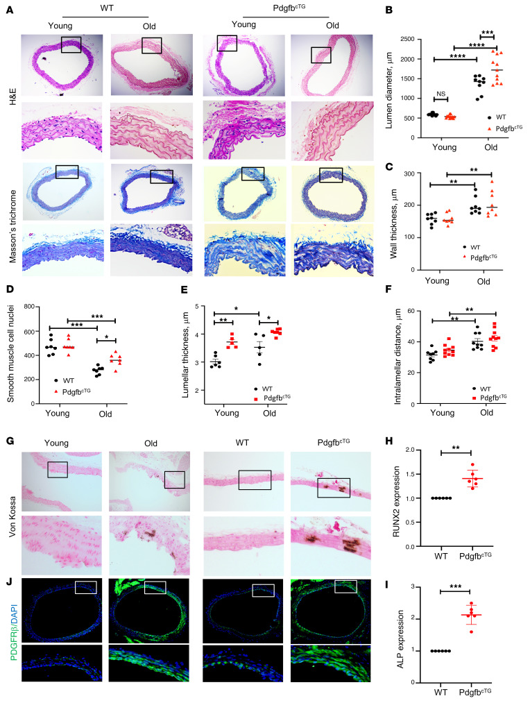
Figure 8. Conditional Pdgfb transgenic mice develop pathological aortic morphology and vascular calcification.
(A) Representative histological staining analysis (10×; inset 40×) showing H&E and Masson’s trichrome staining of aorta from 4- and 18-month-old PdgfbcTG and WT littermates. (B) Lumen diameter, (C) vessel wall thickness, and (D) smooth muscle cell nuclei, Lamellar thickness (E), and intralamellar distance (F) in aortas were calculated. n = 5 to 11. *P < 0.05, **P < 0.01, ***P < 0.001, and ****P < 0.0001 as determined by 1-way ANOVA with Bonferroni post hoc test. (G) Representative micrographs of von Kossa stained sections of the thoracic aorta from young (4-month-old) and old (20-month-old) mice (left panels) and from 6-month-old PdgfbcTG and WT littermates (right panels). (H and I) Aorta tissues were harvested from 6-month-old PdgfbcTG and WT littermates. mRNA expressions of RUNX2 (H) and alkaline phosphatase (ALP) (I) were measured by qRT-PCR. n = 6. Data are mean ± SD, **P < 0.01, ***P < 0.001 as determined by Student’s t tests. (J) Immunofluorescence staining of aortic tissue sections with antibody against PDGFRβ from young (4-month-old) and old (20-month-old) mice (left panels) and from 6-month-old PdgfbcTG and WT littermates (right panels).

