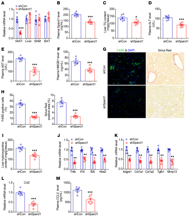
Figure 7. Knockdown of Sparcl1 in iWAT improves NASH pathogenesis in mice.
(A–M) C57BL/6J mice were fed an HFHC diet for 28 weeks, starting at 8 week of age. Then, 5 × 10¹⁰ pfu of either shCon (AAV-scrambled shRNA) or shSparcl1 (AAV-Sparcl1 shRNA) was injected bilaterally into the inguinal fat pads of mice for 4 weeks. n = 6 per group. (A) Relative mRNA levels of Sparcl1 in the iWAT, liver, SKM, and BAT from the 2 groups of mice. (B) Plasma Sparcl1 concentrations. (C) Liver triglyceride (TG) content. (D–F) Plasma levels of ALT (D), AST (E), and HMGB1 (F). (G) F4/80 and Sirius red staining of liver sections. (H) Quantification of the F4/80 and Sirius red staining images. (I) Liver hydroxyproline content. (J and K) Relative mRNA levels of genes involved in liver inflammation and fibrosis. (L) Relative mRNA levels of CCL2 in the livers from the 2 groups of mice. (M) Plasma concentrations of CCL2 from the 2 groups of mice. Data are represented as mean ± SEM. *P < 0.05, **P < 0.01, ***P < 0.001 by 2-tailed Student’s t test (A–F and H–M). Original magnification, ×200 (G).

