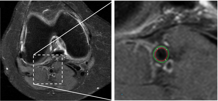
Fig. 1.
Axial MR image of the knee used for assessment of the popliteal vessel wall thickness
The right image shows an enlargement of the popliteal artery area. The green line indicates the outer vessel wall boundary, the red line indicates the lumen. At each analysed slice, the vessel wall thickness is calculated as the average distance between the outer and inner vessel boundary (white lines).

