Figure 1.

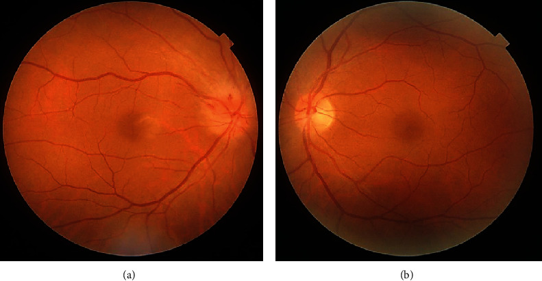
Photographs of the right (a) and left (b) fundus on initial examination. Note the diffuse optic disc swelling in the right eye, which was prominent above the optic disc.

Photographs of the right (a) and left (b) fundus on initial examination. Note the diffuse optic disc swelling in the right eye, which was prominent above the optic disc.