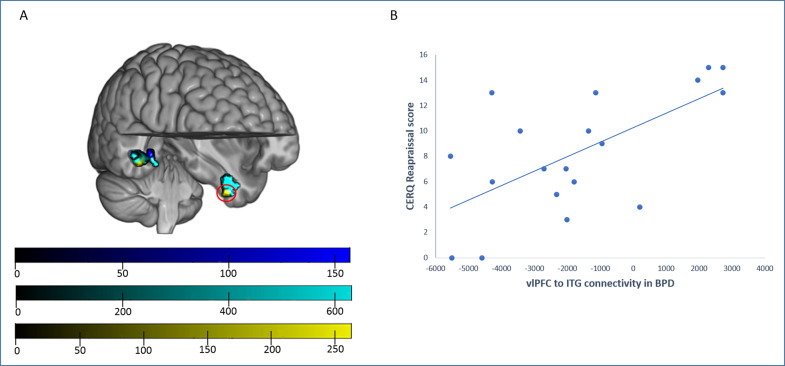
Figure 2.
Between-group differences in generalized psychophysiological interaction (gPPI) analyses from the right vlPFC seed. (A) In comparison to healthy controls (HCs), patients with major depressive disorder (MDD; blue) showed a decreased connectivity with right posterior temporal areas, involving the medial temporal gyrus and the parahippocampal gyrus. Patients with borderline personality disorder (BPD; cyan) showed also decreased connectivity with posterior temporal areas (in this case, with peak differences in the fusiform gyrus) and, specific to these subjects, with the left inferior temporal gyrus (ITG). Moreover, patients with BPD showed, in comparison to the MDD group (yellow), decreased connectivity between the right vlPFC and the left ITG and the right fusiform gyrus. Top color bar: MDD versus HC TFCE (Threshold Free Cluster Enhancement) values; middle color bar; BPD versus HC TFCE (Threshold Free Cluster Enhancement) values; bottom color bar; BPD versus MDD TFCE (Threshold Free Cluster Enhancement) values. (B) The Cognitive Emotion Regulation Questionnaire reappraisal scores correlated positively with right vlPFC–left ITG connectivity (red circle) in the BPD group.

