Figure 2.
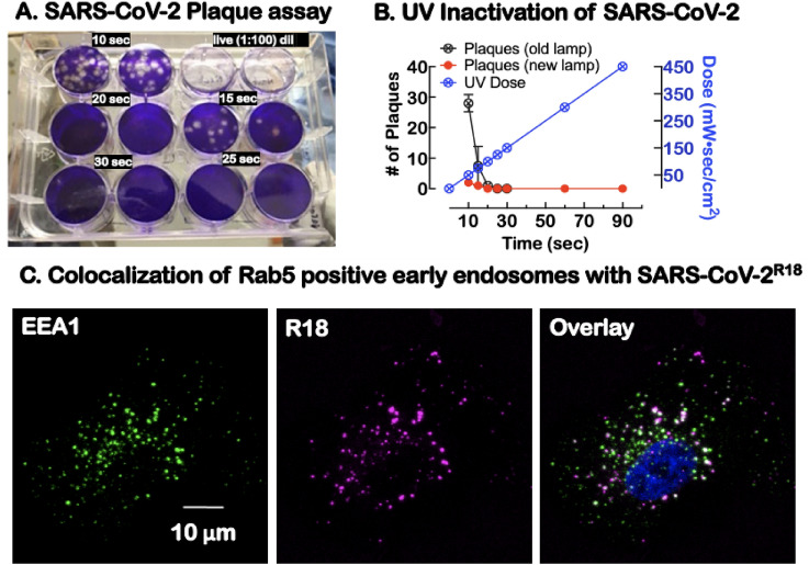
Characterization of UV-inactivated virus for Sars-Cov-2 studies. (A) Duplicate plaque assays of supernatants of Sars-CoV-2 exposed to increasing doses of 254 nm radiation and then tested for viability. The live virus completely lysed the cells at 1:100 dilution relative to UV exposed virions. (B) Graph shows UV dose–response, leading to a significant decrease in plaque-forming units at different doses. For our experiments, a 90 s (450 mW s/cm2) UV dose was used to inactivate the virus before removal from the BSL-3 laboratory. (C) Confocal microscopy imaging of cells after incubation with SARS-CoV-2R18 (magenta) for 15 min, then fixed and labeled for early endosome marker, early endosome antigen 1 or EEA1 (green), an effector protein for Rab5, and nuclei (Hoechst 33,258, blue). SARS-CoV-2R18 vesicles are trafficked to the perinuclear region, and a subset is co-localized with EEA1. Images are maximum projections and have been brightness and contrast-enhanced.

