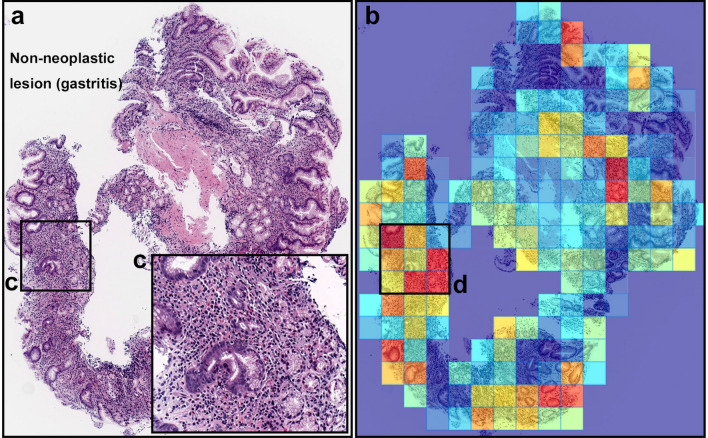
Figure 4.
A representative example of diffuse-type ADC false-positive prediction outputs. (a) is a non-neoplastic lesion (chronic gastritis). Heatmap images (b) exhibited false positive predictions of diffuse-type ADC. The inflammatory tissue with plasma cell infiltration (c) is the possible main cause of false positive (d) due to its analogous nuclear and cellular morphology to diffuse-type ADC cells. Model applied at 20, where the 224 224px heatmap square represents 112 112 m.

