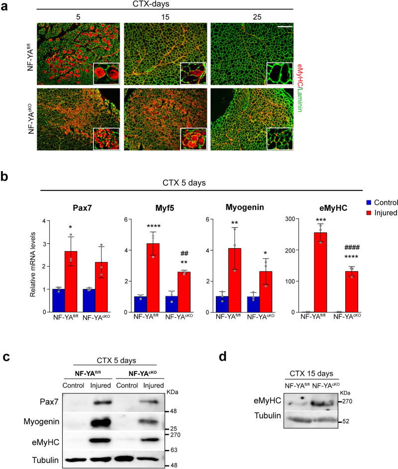
Fig. 2. Loss of NF-YA delays regenerative myogenesis in vivo.
a Representative immunostaining for eMyHC (red) and laminin (green) on TA muscle sections of NF-YAfl/fl and NF-YAcKO mice 5, 15 and 25 days after CTX injury. Nuclei were identified by DAPI staining. Scale bar: 100 µm. b Relative mRNA levels of myogenic markers Pax7, Myf5, Myogenin, eMyHC in whole 5-days injured TA muscles of NF-YAfl/fl and NF-YAcKO mice measured by RT-qPCR. Transcript levels of NF-YAfl/fl and NF-YAcKO uninjured mice have been arbitrarily set at 1. Data represent mean ± s.d. (one-Way Anova: Pax7 F(3,8)=9.66, p = 0.0049; Myf5 F(3,8)=44.63, p < 0.0001; MyoG F(3,8)=9,99, p = 0.0044; eMyHC F(3,8)=170.8, p < 0.0001; n = 3 mice per group. *p < 0.05, **p < 0.01, ***p < 0.001, ****p < 0.0001 vs control; ##p < 0.01, ####p < 0.0001 vs NF-YAfl/fl). c Western blot analysis performed on whole protein extracts of uninjured (control) and 5-days injured TA muscles of NF-YAfl/fl and NF-YAcKO mice. Immunoblots represent protein levels of Pax7, eMyHC, Myogenin and Tubulin. n = 3 experiments. d Western blot analysis of eMyHC on whole protein extracts of 15-days injured TA muscles of NF-YAfl/fl and NF-YAcKO. Tubulin was used as loading control. n = 3 experiments.

