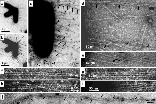
Figure 2.
Multiple bundling of type 1 pili on E. coli cells after the THz irradiation. (a) Three aggregated bacterial cells are shown, one of which carries numerous type 1 pili: these pili have a diameter of 7.0–7.5 nm and vary in length from 0.1 to 2 µm, with maximal length in rare case reaching 3 µm. (b) The copy of (a) in which pili are indicated by black lines. (c) A magnified part of (a) with numerous examples of bundled pili (indicated by arrows) at low magnification. (d) A magnified part of (a) in which the arrows indicate two-pilus bundles. (e) Changes in the diameter of a pilus, indicated by interconnected arrows. (f–i) Examples of 2-, 3-, and 4-pilus bundles. (j) Adhesion of two pili throughout their considerable length. The scale bar is 1 µm in (a,b), 0.1 µm in (c,j), and 50 nm in (d–j).

