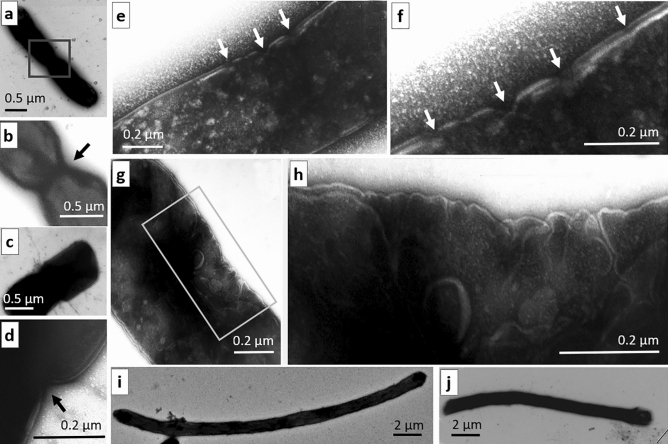
Figure 3.
Disrupted structural organization of the envelope of dividing E. coli cells, as detected after exposure to THz radiation. (a–d) Examples of two dividing bacterial cells at low and high magnification with V-shaped invaginations in the envelope (indicated by arrows) in control. (e,f) Multiple V-shaped invaginations and breaches in the envelope of irradiated bacteria at low and high magnification (indicated by arrows). (g,h) Asymmetric unilateral formation of numerous folds in the envelope in the central part of an irradiated cell. The scale bar is 0.5 μm in (a–c); 0.2 μm in (d–h); and 2 μm in (i,j).

