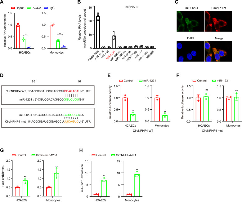
Fig. 6. CircNPHP4 acted as a miRNAs sponge for miR-1231.
A RIP was performed using an antibody against AGO2 on extracts from monocytes and HCAECs. B CircNPHP4 was performed using a circNPHP4-specific probe and control probe in HCAECs. The enrichment of circNPHP4 and microRNAs were detected by qRT-PCR and normalized to the control probe. C Co-localization between circNPHP4 and miR-1231 was observed by RNA in situ hybridization in HCAECs. Nuclei were stained with DAPI. Scale bar = 20 µm. D Schematic showing the predicted miR-1231 sites in circNPHP4. A Luciferase assay where monocytes were co-transfected with a scrambled control, miR-1231 mimic, and a luciferase reporter plasmid containing wild-type circNPHP4 (circNPHP4-WT) (E) or mutant circNPHP4 (circNPHP4-mut) (F). G qRT-PCR showed the level of circNPHP4 in the streptavidin-captured fractions from the monocytes and HCAECs lysates after transfection with biotinylated miR-1231 or control RNA. H Expression of miR-1231 was analyzed using qRT-PCR following circNPHP4 knockdown. Data are presented as means ± SD; a significant difference was identified with Student’s t test. *P < 0.05; **P < 0.01; ns (not significant).

