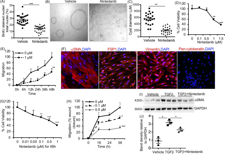
Fig. 1. Effect of Nintedanib treatment on ADPKD renal cystic epithelial cells and myofibroblasts.
A BrdU-incorporation assay in human ADPKD renal cystic epithelial cells treated with nintedanib (1.5 µM for 24 h). n = 4 biological replicates (patient samples) and seven technical replicates each. B Images of microcyst growth in 3D collagen matrix by human ADPKD renal cystic epithelial cells treated with vehicle or nintedanib (1.5 µM) for 12 days. 1X magnification, and (C) quantitation of cyst diameter. n = 4 biological replicates and 5 or 6 technical replicates each. D Human ADPKD renal myofibroblasts treated with nintedanib for 48 h. MTT assay and E migration (% wound closure in a scratch assay) in the presence of 0 or 1 µM nintedanib. n = 4 biological replicates and three technical replicates each for D and E. F Human renal ADPDK myofibroblasts immunostained for αSMA, FSP1, vimentin, or pan-cytokeratin. G NRK-49F rat renal fibroblasts treated with nintedanib for 48 h. MTT assay. n = 7. H Migration of NRK-49F cells treated with nintedanib n = 4. I Immunoblot of NRK-49F cells incubated with TGFβ (2 ng/ml) and nintedanib (1.5 μM) for 48 h and (J) quantitation of band density. *P < 0.05 **P < 0.01, ***P < 0.001, vs vehicle or 0 µM nintedanib.

