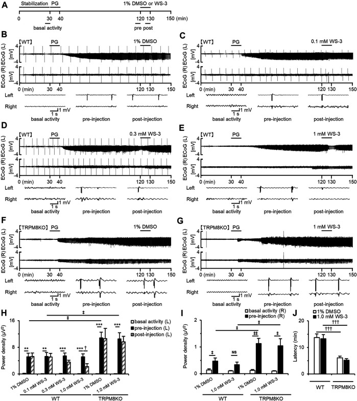
FIGURE 2.
Suppressive effects of WS-3 on PG-induced EDs in WT mice and deterioration of EDs due to lack of TRPM8 channels. (A) Experimental protocol. Mice were anesthetized with urethane. The power of the low beta band in ECoG was integrated for 5 min during the basal activity (25–30 min) and the pre- (115–120 min) and post- (125–130 min) injection periods. Examples of changes in EDs with (B) PG + 1% DMSO (n = 6), (C) PG + 0.1 mM WS-3 (n = 5), (D) PG + 0.3 mM WS-3 (n = 6), and (E) PG + 1.0 mM WS-3 (n = 6) in WT mice, and (F) PG + 1% DMSO (n = 6) and (G) PG + 1.0 mM WS-3 (n = 6) in TRPM8KO mice. (B–G) Representative ECoG traces for 5 s during basal activity and the pre- and post-injection period. (H) The integrated value of the low beta bands in the affected (Light) side during basal activity (white) and the pre- (black) and post-injection (grey-oblique line) periods. (I) The integrated value of the low beta bands in the contralateral (Right) side during basal activity (white) and the pre-injection (black) period. (J) Latency of EDs evoked in WT (white) and TRPM8KO mice (grey). The results are shown as mean ± SEM; **p < 0.01, ***p < 0.001, Tukey’s test; † p < 0.05, ††† p < 0.001, Student’s t-test; ‡ p < 0.05, ‡‡ p < 0.01, Welch’s t-test. NS, no significant.

