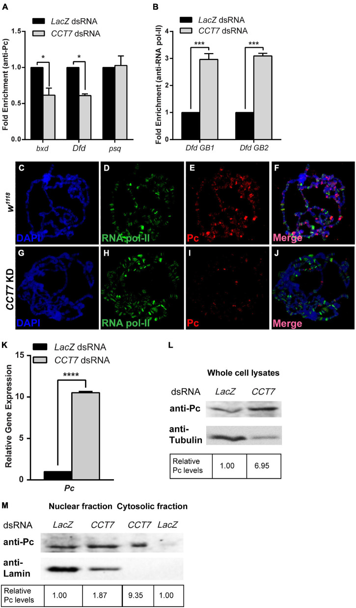
FIGURE 4.
Chaperonin containing TCP-1 subunit 7 (CCT7) is required for association of Polycomb (Pc) at chromatin. (A) Chromatin immunoprecipitation (ChIP) using anti-Pc antibody from CCT7 knockdown cells showed decreased enrichment of Pc at bxd and Dfd as compared to LacZ dsRNA-treated control cells. ChIP was analyzed using fold enrichment over input. Error bars represent SEM for two independent experiments. Statistical significance was calculated using two-way ANOVA (* p ≤ 0.01). (B) ChIP from cells treated with dsRNA against CCT7 showed increased RNA pol-II along the gene body of Dfd (labeled Dfd GB1 and Dfd GB2) as compared to control correlating with an increased expression of Dfd (Figure 2E) in CCT7-depleted cells. ChIP was analyzed using fold enrichment over input. Error bars represent SEM for two independent experiments. Statistical significance was calculated using two-way ANOVA (*** p ≤ 0.001). (C–J) Polytene chromosomes prepared from w1118 and CCT7-depleted salivary glands and stained with anti-RNA pol-II and anti-Pc antibodies. As compared to RNA pol-II staining used as a positive control (D,H), a strongly diminished binding of Pc was observed after depletion of CCT7 (I) when compared with w1118 control (E). (K,L) Drastic increase in expression of Pc in CCT7-depleted cells. CCT7-depleted cells displayed significantly higher expression of Pc in real-time PCR analysis (K) as well as on Western blot (L) when compared to control cells. Error bars represent SEM for two independent experiments. Statistical significance was calculated using t-test (**** p ≤ 0.0001). (M) Western blot probed with anti-Pc and anti-Lamin antibodies showing nuclear and cytosolic fractions isolated from CCT7-depleted and control cells. Besides its presence in the nucleus, Pc is visible as enriched in the cytosolic fraction of CCT7-depleted cells as compared to the cytosolic fraction of LacZ dsRNA-treated control. The same blot probed with anti-Lamin served as a control for purity of nuclear and cytosolic fractions. Western blots were quantified using ImageJ software, and data were normalized using LacZ dsRNA-treated cells as control.

