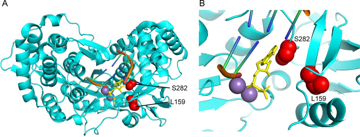
Fig. 6.
Structural insights into sofosbuvir resistance mutations found in hepatitis C virus NS5B (4WTJ). (A) Overview of NS5B structure with template and elongating RNA strands (orange), incoming ADP (yellow), manganese ions (purple), and two amino acid residues for which sofosbuvir resistance mutations exist for (red, S282T, L259F). (B) Zoomed view of the spatial position of S282, L159, and the incoming ADP nucleotide. As suggested by Appleby et al., due to the proximity of residue 282 to incoming nucleotides, the T282 mutant may sterically clash with the 2′ methyl group found on the functional sofosbuvir molecule, prohibiting incorporation [117]. Structures oriented in PyMol, PDB: 4WTJ.

