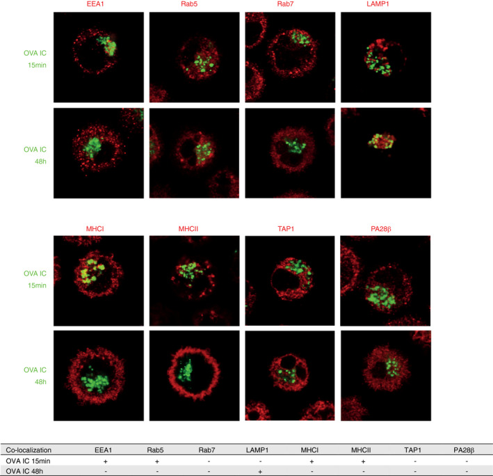
FIGURE 2.

Characterization of the antigen storage compartments in DCs upon FcγR targeting. DCs were pulsed with OVA IC (Alexa Fluor 488, green) for 15 min or pulsed with OVA IC for 2 h and chased for 48 h. OVA IC presence in DCs was imaged by confocal microscopy, and DIC was used to image cell contrast. Specific antibodies against EEA1, Rab5, Rab7, LAMP1, MHCI, MHCII, TAP1 and PA28β (red) were used and analysed for co‐localization with OVA IC. Co‐localization between OVA IC and the antibodies is summarized in a table, ‘+’ indicates co‐localization, ‘‐’ indicates no co‐localization
