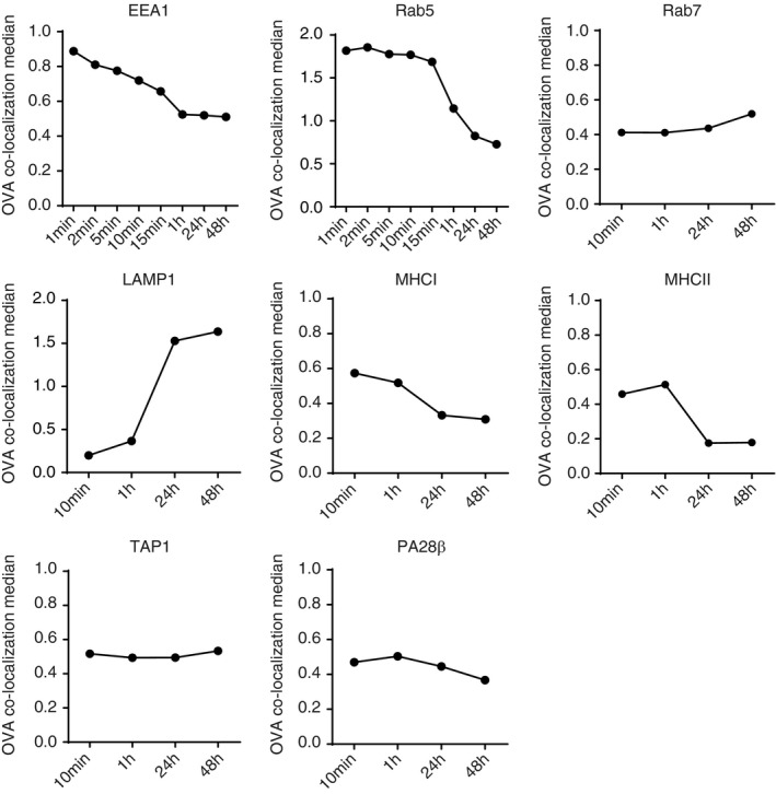
FIGURE 3.

Characterization of the antigen storage compartments in DCs with imaging flow cytometry. DCs were pulsed with OVA IC (Alexa Fluor 647 labelled OVA) for 1, 2, 5, 10, 15 min or 1 h (different for each antibody), and pulsed for 2 h and chased for 24 and 48 h. Co‐localization between OVA IC and EEA1, Rab5, Rab7, LAMP1, MHCI, MHCII, TAP1 and PA28β was analysed by imaging flow cytometry
