FIGURE 4.

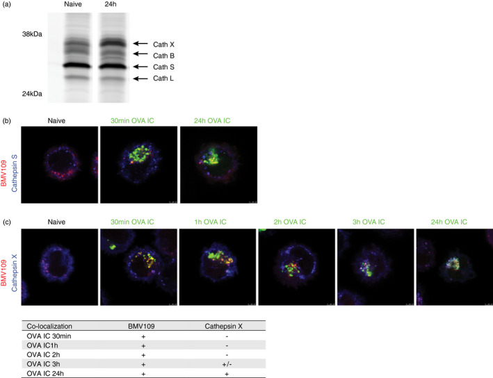
The presence of cathepsins in the antigen storage compartments in DCs. Naïve DCs or DCs pulsed with OVA IC for 2 h and chased for 24 h were run on a 15% PAGE gel. Quenched activity‐based probe BMV109 was used to stain active cathepsin X, B, S and L, indicated by arrows (a). Co‐localization between BMV109 (red) and specific antibody against cathepsin S (blue) was analysed in naïve DCs with confocal microscopy. DCs were pulsed with OVA IC (Alexa Fluor 488) for 30 min or pulsed for 2 h and chased for 24 h. Co‐localization between OVA IC (green), cathepsin S (blue) and BMV109 (red) was analysed by confocal microscopy (b). Naïve DCs, DCs pulsed with OVA IC (Alexa Fluor 488) for 30 min, 1, 2, 3 h or DCs pulsed for 2 h and chased for 24 h were stained with cathepsin X antibody and BMV109. Co‐localization between OVA IC (green), cathepsin X (blue) and BMV109 (red) was analysed by confocal microscopy. Co‐localization between OVA IC, BMV109, cathepsin X and cathepsin S is summarized in a table, ‘+’ indicates co‐localization, ‘+/−’ indicates partial co‐localization, and ‘‐’ indicates no co‐localization (c)
