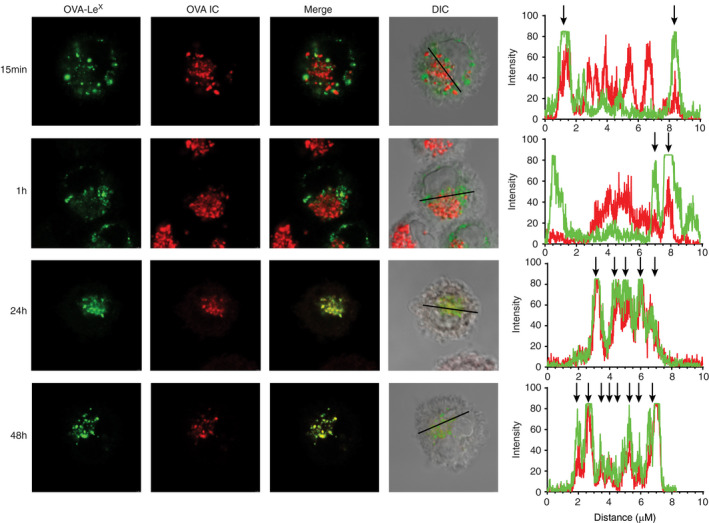
FIGURE 6.

Antigens targeted to FcγRs and MGL1 on DCs end up in the same compartments. DCs were pulsed with OVA IC (Alexa Fluor 647) or OVA‐LeX (DyLight 488) for 15 min and 1 h, or pulsed for 2 h and chased for 24 or 48 h. Co‐localization between OVA IC (red) and OVA‐LeX (green) was visualized by confocal microscopy and DIC was used to image cell contrast. Histograms for each fluorophore were created for a selected area (indicated by a line on the image) and overlays were made with the ImageJ software. Arrows indicate co‐localization between OVA IC (red) and OVA‐LeX (green)
