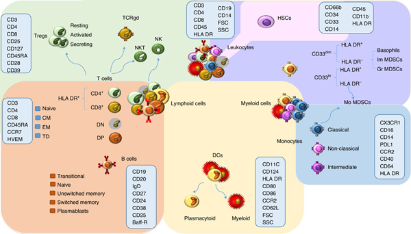
Fig. 1 |. Schematic representation of the main leukocyte subsets assessed by GWAS.
The background colors indicate different cell groupings: T-cell populations are indicated in green, B cells in orange, DCs in yellow, monocytes in blue, other myeloid cells in violet, and hematopoietic stem cells in pink. The markers assessed for MFI are indicated within the light blue rectangles. TCRgd, gamma delta T cells; NK, natural killer cells; NKT, NK T cells; DN, CD4− CD8− T cells; DP, CD4+ CD8+ T cells; CM, central memory; EM, effector memory; HSCs, hematopoietic stem cells; Im MDSCs, immature myeloid-derived suppressor cells; Gr MDSCs, granulocytic MDSCs; Mo MDSCs, monocytic MDSCs; FSC, forward scatter; SSC, side scatter.

