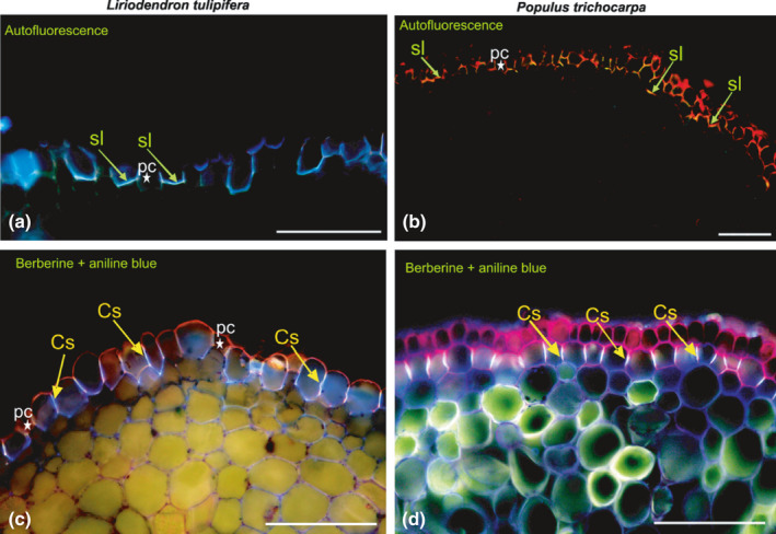
Fig. 18.

Visualisation of exodermis in Liriodendron tulipifera and Populus trichocarpa roots; exodermis autofluorescence (a, b) and root sections stained with berberine and aniline blue (c, d). Cs, Casparian strips; sl, suberin lamellae; pc, passage cells (asterisks). Bars, 100 µm.
