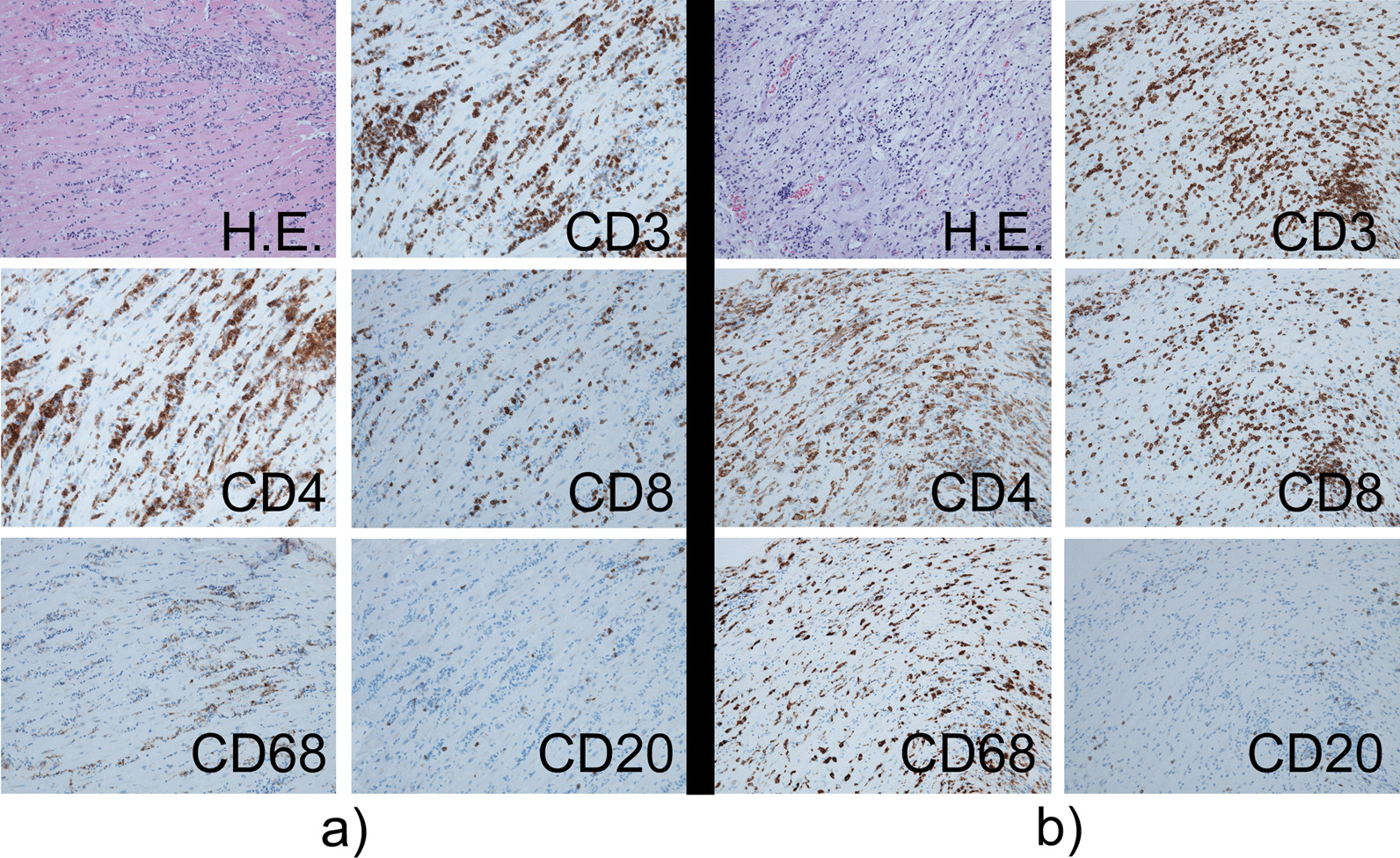
Fig. 5.

Photomicrographs of the endomyocardial biopsy sample before and after treatment. Hematoxylin–eosin staining and immunohistochemical staining of sections of the interventricular septum demonstrating staining with anti-CD3, anti-CD4, anti-CD8, anti-CD68, and anti-CD20 antibodies. All images are displayed at 20× the original magnification. Histologic findings show lymphocytic infiltration in the myocardium comprising CD3 positive T lymphocytes, many of which were positive for CD4 compared with CD8, abundance of CD68-positive macrophage infiltrate, and only rare B lymphocytes before treatment (a). Despite intense treatment, the remaining prominent inflammatory infiltrate consisted of CD3+, CD4+, and CD8+ T cells, and CD68+ macrophages, suggesting smoldering myocarditis (b)
