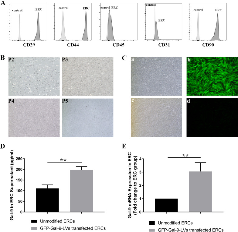
Fig. 1.
Characterization of ERCs and upregulation of Gal-9 in ERCs. A Cell surface markers of ERCs were measured by flow cytometry. ERCs express CD29, CD44, CD90, while lacking of CD45, CD31. B Cell morphology images (magnification 40×) of ERCs at P2-5. C Brightfield and fluorescence pictures are shown on the left and right side. Representative picture of GFP-Gal-9-LVs transfected ERCs (a-b) and unmodified ERCs(c-d) (magnification 40×). D Gal-9 protein expression in unmodified ERCs and GFP-Gal-9-LVs transfected ERCs. n = 3. **p < 0.01. E Gal-9 mRNA relative expression in unmodified ERCs and GFP-Gal-9-LVs transfected ERCs. n = 3. **p < 0.01. p value was calculated using unpaired t-test. Abbreviations: ERCs endometrial regenerative cells, Gal-9 galectin-9, P3-5 passage3-5

