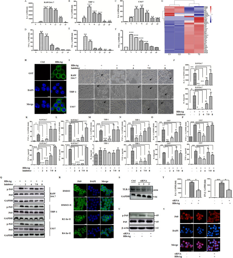
Fig. 1.
HBeAg regulated the expression of multiple TLRs in macrophages, while the inflammatory response of HBeAg-induced macrophages was inhibited by TLRs. RAW 264.7 macrophages were stimulated with HBeAg (2 μg/ml) at different time points, and then the expression of IL-6 and TNF-α was detected respectively using qRT-PCR (A, D). THP-1 (B, E) and U937 cells (C, F) were pretreated with PMA at a final concentration of 50 ng/ml. After replacing the fresh medium, they were stimulated with HBeAg (2 μg/ml) at different time points, and the expression of IL-6 and TNF-α was detected respectively using qRT-PCR. RAW264.7 macrophages were stimulated with HBeAg for 24 h, and RNA sequencing assay was performed. The expression profile of growth factors, cytokines, chemokines, and TLRs were selected and displayed (G). H After incubation with HBeAg for 1 h at 4 °C, RAW 264.7 macrophages were fixed with 1% paraformaldehyde for 20 min at room temperature. After blocking, cells were incubated with Alexa Flour 488-conjugated primary antibody for 1 h at 37 °C followed by DAPI for nuclear staining, then captured using a confocal fluorescence microscopy. RAW 264.7 macrophages and PMA-pretreated THP-1/U937 cells were treated with DMSO or inhibitors of TLR-2, 4, 8, 7/9 (10μΜ) for 30 min. The cell morphology was analyzed after HBeAg treatment for 24 h (I). The arrows indicate activated macrophages. RAW 264.7 macrophages were firstly treated with DMSO or inhibitors of TLR-2, 4, 8, 7/9 for 30 min, and then they were treated with HBeAg for 4 h. The expression and secretion of IL-6, TNF-α, and IFN-β were tested by qRT-PCR and ELISA respectively (J–L). PMA-pretreated THP-1 (M–N) and U937 cells (O–P) were treated with DMSO or inhibitors of TLR-2, 4, 8, 7/9 for 30 min, and then they were treated with HBeAg for 4 h. The expression and secretion of IL-6 and TNF-α were tested by qRT-PCR and ELISA respectively. Macrophages, including RAW264.7, PMA-pretreated THP-1 and U937 cells, were firstly treated with DMSO or inhibitors of TLR-2, 4, 8, 7/9 (10μΜ) for 30 min, then they were treated with HBeAg for another 20 min. The phosphorylation level of p65 was tested by western blot assay (Q). Additionally, the effect of TLR-2/4 inhibitors on the nuclear translocation of p65 was determined (R). The siRNAs for TLR-3 or negative control were transfected into RAW264.7 macrophages for 48 h, and then the cells were treated with HBeAg for 4 h. The expression of TLR-3 was tested by western blot assay (S), and the level of IL-6 and IFN-β (T) was tested by qRT-PCR. The siRNAs for TLR-3 or negative control were transfected into RAW264.7 macrophages for 48 h, and then the cells were treated with HBeAg for 20 min. The phosphorylation level and nuclear translocation of p65 were tested separately (U-V). *P < 0.05, **P < 0.01, ***P < 0.001

