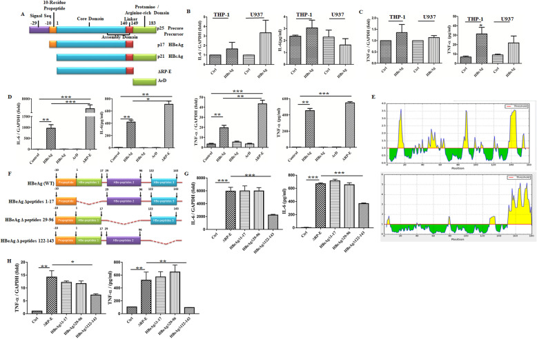
Fig. 3.
The C-terminal peptides of HBeAg played a key role in macrophage activation. Comparison of the structure for HBeAg, HBcAg, and their precore precursor proteins (A). PMA-pretreated THP-1/U937 cells were treated with 2 μg/ml HBcAg for 4 h, then the expression and secretion of IL-6 and TNF-α were tested by qRT-PCR and ELISA respectively (B, C). RAW264.7 macrophages were stimulated with HBeAg, HBcAg, ArD, and ΔRP-E (2 μg/ml) for 4 h respectively, and then the expression and secretion of IL-6 and TNF-α were tested by qRT-PCR and ELISA (D). Bioinformatics analysis of the surface accessible peptides for HBeAg (upper part) and HBcAg (lower part) was predicted (E). Schematic illustration of wild-type and truncation mutants of HBeAg (F). RAW264.7 macrophages were stimulated with ΔRP-E and truncation mutants of HBeAg (2 μg/ml) based on the analysis of the surface accessible peptides, then the expression and secretion of IL-6 and TNF-α were tested by qRT-PCR and ELISA (G, H). *P < 0.05, **P < 0.01, ***P < 0.001

