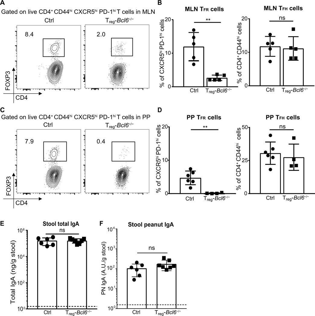
Fig. 6. Production of PN IgA is independent of TFR cells.
(A and C) Representative flow plot and (B and D) frequencies of TFR and TFH cells in (A and B) MLN and (C and D) PP from control (Bcl6flox/flox) or Treg-Bcl6−/− (Foxp3creBcl6flox/flox) littermates 8 days after one PN + CT immunization. TFR cells (FOXP3+) were gated on TFH cells (CXCR5hi PD1hi). TFH cells were gated on CD4+ CD44hi live activated T cells (TCRβ+). (E and F) ELISA quantification of total IgA and PN IgA in stool of Treg-Bcl6+/+ or Treg-Bcl6−/− littermates 1 week after sixth immunization with PN + CT. Figures represent data from three independent experiments with three to six mice per group. Median of each group was compared with Mann-Whitney U test whereby (**) indicates significant differences with P <0.01. Error bars indicate SD

