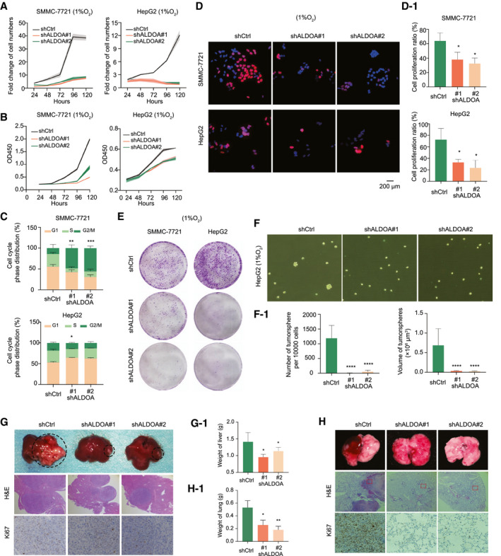
Fig. 2.

ALDOA promoted HCC cell proliferation and migration under hypoxia in vitro and in vivo. (A,B) Knockdown of ALDOA significantly reduced numbers (A) and growth (B) of SMMC‐7721 cells and HepG‐2 cells under hypoxia. (C) Knockdown of ALDOA induced HCC cell cycle arrest under hypoxia. (D) Knockdown of ALDOA significantly reduced HCC cell proliferation under hypoxia by EdU assay. Quantification of fold change is shown in D‐1. (E) Knockdown of ALDOA impaired colony formation in HCC cells under hypoxia. (F) Knockdown of ALDOA impaired mammosphere formation of HepG2 cells under hypoxia. Quantification of fold change is shown in F‐1. (G) Knockdown of ALDOA dramatically suppressed liver tumor growth in orthotopic xenograft mouse models. Stable ALDOA‐knockdown Hepa1‐6 cells (5 × 10⁶), and control cells were injected into the liver of each female C57 mouse. Three weeks after injection, livers were separated for pathological analysis. Liver weights are shown in G‐1. (H) Knockdown of ALDOA abolished lung metastasis from HCC. Hepa1‐6 cells (5 × 10⁶) were tail vein–injected into female C57 mice. Formation of metastatic foci in the lung was pictured after 2 weeks. Metastases were confirmed by pathological analysis. Lung weights are shown in H‐1. *P < 0.05, **P < 0.01, ***P < 0.001, ****P < 0.0001. Abbreviations: H&E, hematoxylin and eosin; OD, optical density.
