Fig. 6.

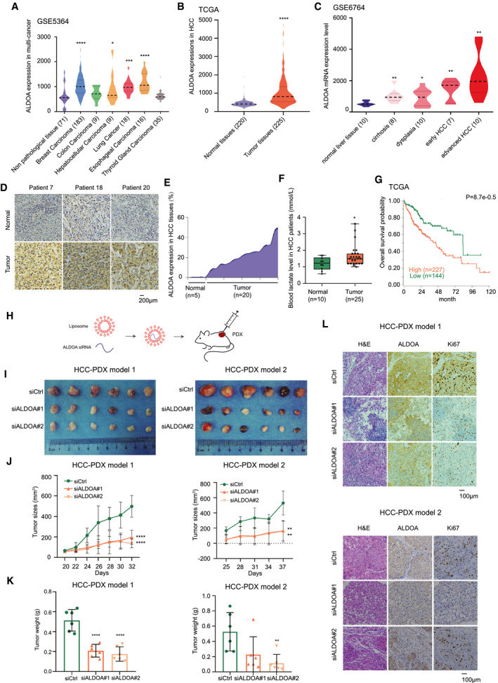
Clinical significance of ALDOA inhibition in HCC. (A) ALDOA expression in multiple tumors. (B) Up‐regulation of ALDOA in HCC tissues compared with normal liver tissues. (C) The expression level of ALDOA was correlated with pathological stages of HCC. (D) ALDOA expression level in clinical patients and their pericarcinomatous normal tissues by IHC assay. (E) Quantification of ALDOA expression in HCC tissues and normal liver tissues from IHC results. (F) Levels of blood lactate acid in patients with clinical HCC compared to healthy people. (G) Prognostic significance of ALDOA up‐regulation in HCC. (H) Schematic diagram of targeting ALDOA by siRNAs in vivo. (I) Inhibition of ALDOA by RNAi in two HCC‐PDXs. (J,K) Tumor growth curve (I) and tumor weight (J) of HCC‐PDXs treated with two in vivo–optimized siRNAs. (L) Hematoxylin and eosin staining and IHC images of ALDOA and Ki67 in randomly selected HCC‐PDX#1 and HCC‐PDX#2 tumors. *P < 0.05, **P < 0.01, ***P < 0.001, ****P < 0.0001. Abbreviation: H&E, hematoxylin and eosin.
