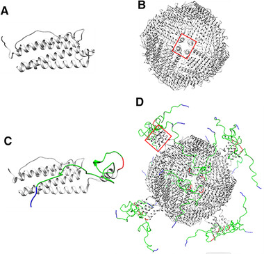
FIGURE 6.

Schematic illustration of assumed protein structures. (A) “flip” HFn subunit. (B) “flip” HFn assembly. (C) “flop” modified subunit (HFn‐PAS‐RGDK). (D) “flop” assembly. Light grey parts represent HFn, red parts represent enzyme‐cleavable sequence GFLG, green parts are PAS peptides and blue parts are RGDK peptides. Structure of HFn was originally obtained from PDB (5N27) and then modified and visualized using Chimera. [27] Red rectangle indicates E‐helix location
