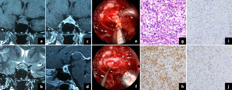
Figure 2.
Magnetic resonance imaging showed a 1.7×1.2 cm cystic lesion with a thickened pituitary stalk and mainly peripheral enhancement (A–D). Intraoperative pictures revealed a soft yellow lesion (E) with pus-like fluid (F). Pathologic examination showed that the pituitary gland was infiltrated by foamy histocytes and lymphocytes (G, ×200). The foamy cells were immunopositive for CD68 (H, ×200) and immunonegative for CD1a (I, ×200) and S-100 protein (J, ×200).

