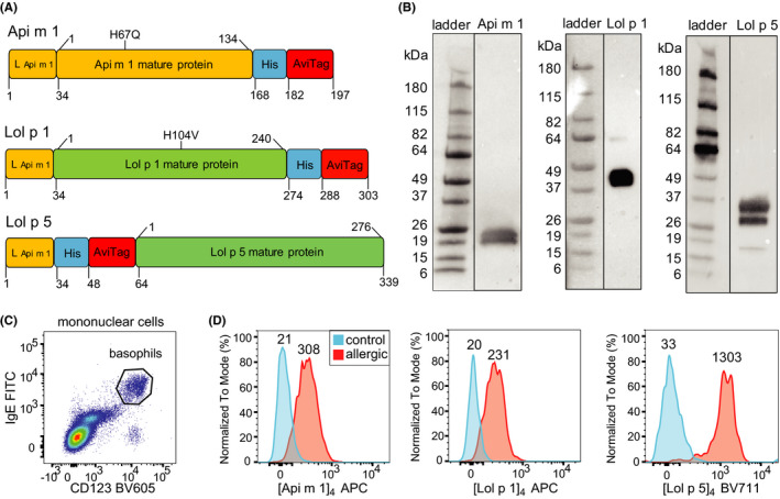
FIGURE 1.

Detection of allergen sensitization by quantifying binding of fluorescent allergen components of BV and RGP to basophils. A) Schematic diagram of DNA constructs for generation of the recombinant allergens Api m 1, Lol p 1, and Lol p 5. Recombinant allergens contained mutations to render the catalytic site inactive (Api m 1 H67Q, Lol p 1 H104V). Leader peptide and spacer (L); 6‐histidine tag (His); avidin tag (AviTag). B) Western blots of recombinant allergens detected with anti‐His antibody. C) Representative flow plot for detection of circulating basophils (CD123+IgE+) within gated mononuclear cells (Figure S1). D) Histograms depicting representative fluorescent staining of Api m 1, Lol p 1, and Lol p 5 tetramers on basophils from relevant allergic patients (red) and controls (blue). Values above the histograms represent median fluorescence intensity values of the population.
