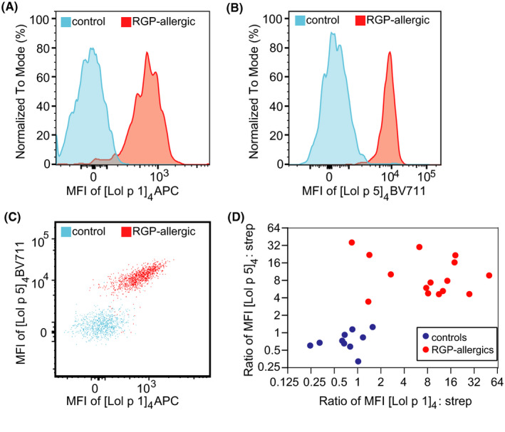
FIGURE 5.

Multiplex flow cytometry using multiple allergen tetramers in a single tube as a component‐resolved diagnostic test for allergy. A, B) Representative histograms of basophils from thawed PBMC of a control (blue) or an RGP‐allergic patient (red) stained with Lol p 1 and Lol p 5 tetramers in a single tube. C) 2D dot plot of Lol p 1 and Lol p 5 tetramer staining of the basophils from the control and patient in panels A and B. D) Scatterplot depicting MFI ratios of allergen:streptavidin for [Lol p 1]4‐APC vs strep‐APC and [Lol p 5]4‐BV711 vs strep‐BV711 on basophils from controls (blue, n = 10) and RGP‐allergic subjects (red, n=15) stained with both allergen tetramers or both streptavidin conjugates in a single tube.
